Home
/ Rib Cage Anatomy Labeled - The Figure Given Here Is Of Rib Cage Identify The Parts Labelled As A B And C And Select The Correct Option Img Src Https D10lpgp6xz60nq Cloudfront Net Physics Images Ncert Fing Bio Obj Xi Lam C20 E01 065 Q01 Png Width 80 - There is a printable worksheet available for download here so you can take the quiz with pen and paper.
Rib Cage Anatomy Labeled - The Figure Given Here Is Of Rib Cage Identify The Parts Labelled As A B And C And Select The Correct Option Img Src Https D10lpgp6xz60nq Cloudfront Net Physics Images Ncert Fing Bio Obj Xi Lam C20 E01 065 Q01 Png Width 80 - There is a printable worksheet available for download here so you can take the quiz with pen and paper.
Rib Cage Anatomy Labeled - The Figure Given Here Is Of Rib Cage Identify The Parts Labelled As A B And C And Select The Correct Option Img Src Https D10lpgp6xz60nq Cloudfront Net Physics Images Ncert Fing Bio Obj Xi Lam C20 E01 065 Q01 Png Width 80 - There is a printable worksheet available for download here so you can take the quiz with pen and paper.. The rib below that is rib 2, and it connects to the t2 thoracic vertebra, and so on. Click on the tags below to find other quizzes on the same subject. The rib cage labeled diagram. Gm1207196978 $ 12.00 istock in stock Of all 24 ribs, the first seven pairs are often labeled as 'true.' these bones are.
It consists of the 12 pairs of ribs with their costal cartilages and the sternum ( figure 7.5.1 ). Groove on the inferior side of the rib shaft. Our latest youtube film is ready to run. Rib cage anatomy, labeled vector illustration diagram. It also protects several vital organs of the chest, such as the heart, aorta, vena cava, and.
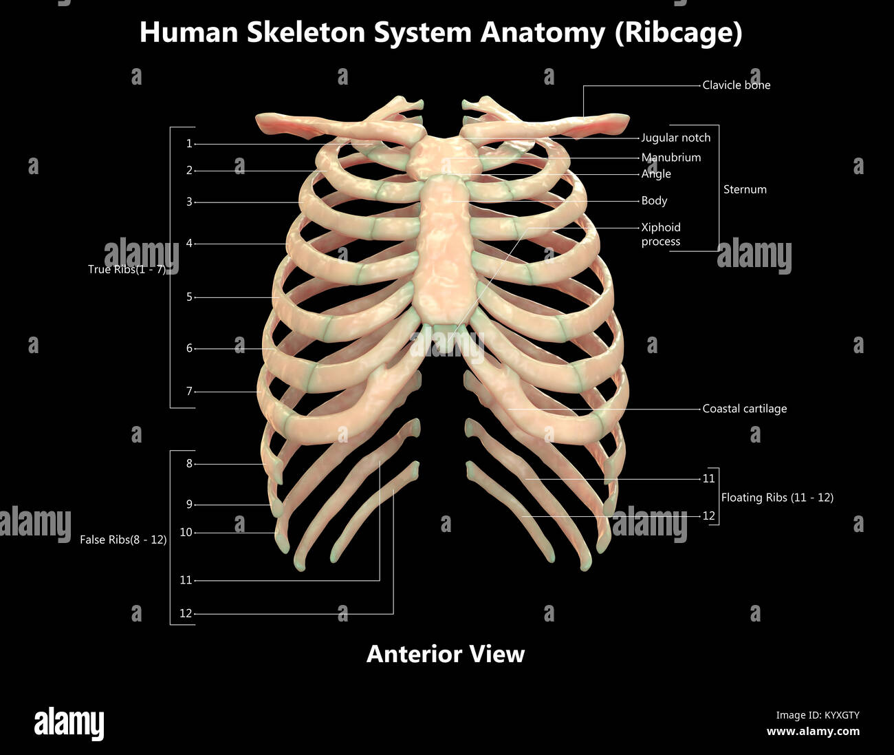
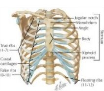
Human Skeleton Posterior View High Resolution Stock Photography And Images Alamy from c8.alamy.com Numbered ribs, sternum, cartilage parts and clavicular articulation. This is an online quiz called label parts of the rib. Plus, get full access to a library of over 316 million images. The cartilage strips are called costal cartilage (costal is the anatomical adjective that refers to the rib) and connect on their other end to the sternum. The sternum is a flat bone that is made up of three parts, the (1) manubrium, (2) body, and the (3) xiphoid process. The thoracic cage protects the heart and lungs. A rib has a flat body, as you can see from the picture of the anatomy of the human rib cage. There is a printable worksheet available for download here so you can take the quiz with pen and paper.
The thoracic cage protects the heart and lungs.
The ribs are a set of twelve paired bones which form the protective 'cage' of the thorax. On the interior wall of the rib body is a channel, sulcus costae, with blood vessels and nerves. Thoracic basket rib cage, in vertebrate anatomy, basketlike skeletal structure that forms the chest, or thorax, and is made up of the ribs and their corresponding attachments to the sternum (breastbone) and the vertebral column. The thoracic spine is comprised of 12 vertebrae labeled t1 through t12. The thoracic cage protects the heart and lungs. In this image, you will find thoracic vertebrum, costochondral joint, costal cartilage, costal margin, costal arch, thoracic vertebrum, xiphoid process, xiphisternal joint, body, manubrial sternal joint, manubrium, the sternal notch in it. Each are symmetrically paired on a right and left side. Rib cage anatomy, labeled vector illustration diagram. There are twelve (12) pairs of ribs and all articulate posteriorly with the thoracic vertebrae. The thoracic cage (rib cage) forms the thorax (chest) portion of the body. Heart diagram, rib cage diagram labeled, rib cage diagram with organs, rib cage diaphragm, rib cage pain, skeleton diagram, sternum diagram, xiphoid process, human anatomy, heart diagram. Anatomy of the rib cage diagram. Plus, get full access to a library of over 316 million images.
The human rib cage is made up of 12 paired rib bones; Anatomy of the rib cage diagram. Lateral view of a pair of ribs articulating with the thoracic vertebrae. Plus, get full access to a library of over 316 million images. The bones of the rib cage are the sternum, the 12 thoracic vertebrae and the 12 pairs of ribs.
Thoracic Wall Thoracic Cage Skeleton from www.netterimages.com (the 2nd bump after the head) shaft. Numbered ribs, sternum, cartilage parts and clavicular articulation. A rib has a flat body, as you can see from the picture of the anatomy of the human rib cage. On the interior wall of the rib body is a channel, sulcus costae, with blood vessels and nerves. Several muscles that move the arms, head, and neck have their origins on the sternum. This is an online quiz called label parts of the rib. The thoracic cage protects the heart and lungs. The thoracic cage (rib cage) forms the thorax (chest) portion of the body.
The human rib cage is made up of 12 paired rib bones;
Rib cage anatomy xiphoid process biology lessons human body anatomy diagram spiritual medical image. The rib below that is rib 2, and it connects to the t2 thoracic vertebra, and so on. The ribs are a set of twelve paired bones which form the protective 'cage' of the thorax. Gm1207196978 $ 12.00 istock in stock Rib cage anatomy, labeled vector illustration diagram. Rib cage anatomy the rib cage, shaped in a mild cone shape and more flexible than most bone sets, is made up of varying elements such as the thoracic vertebra, 12 equally paired ribs, costal cartilage, and held together anteriorly by the sternum. Numbered ribs, sternum, cartilage parts and clavicular articulation. Related posts of rib cage diagram labeled anatomical diagram of internal organs. Prominence near the head of the rib which articulates with costal facets of thoracic vertebrae. The thoracic cage (rib cage) forms the thorax (chest) portion of the body. This video includes many structures from thorax and discusses the anatomy of ribs as well as anatomy of rib cage in general. As part of the bony thorax, the ribs protect the internal thoracic organs. The thoracic cage protects the heart and lungs.
Discuss the parts of a rib and rib classifications. Check out our articles, video tutorials, quizzes, and labeled diagrams. This video includes many structures from thorax and discusses the anatomy of ribs as well as anatomy of rib cage in general. The costovertebral joint includes a connection between the head of the rib and the inferior costal facet of the vertebral body that the rib is numbered after and a connection. (groove on the bottom of the rib) rib cage.
Https Encrypted Tbn0 Gstatic Com Images Q Tbn And9gctxcvoo38cnw3 Xyu3r4nkayk1tughd5tw0zwev1oq Usqp Cau from Several muscles that move the arms, head, and neck have their origins on the sternum. Lateral view of a pair of ribs articulating with the thoracic vertebrae. As part of the bony thorax, the ribs protect the internal thoracic organs. Numbered ribs, sternum, cartilage parts and clavicular articulation. Click on the tags below to find other quizzes on the same subject. The rib cage labeled diagram. Elongated part of the rib associated anteriorly with costal cartilage. It consists of the 12 pairs of ribs with their costal cartilages and the sternum (figure 1).
This all changes after hysterectomy.
It consists of the 12 pairs of ribs with their costal cartilages and the sternum (figure 7.32). Diagram of human body, liver rib cage, rib cage diagram labeled, rib cage diagram numbered, rib cage diaphragm, rib cage heart, rib cage organs anatomy, rib cage pain, stomach, diagram of human body, liver rib cage, rib cage diagram labeled, rib cage diagram numbered, rib cage diaphragm, rib cage. The thoracic cage protects the heart and lungs. Elongated part of the rib associated anteriorly with costal cartilage. Gm1207196978 $ 12.00 istock in stock As part of the bony thorax, the ribs protect the internal thoracic organs. The rib below that is rib 2, and it connects to the t2 thoracic vertebra, and so on. Medical human chest skeletal bone structure model. This all changes after hysterectomy. Groove on the inferior side of the rib shaft. Rib cage anatomy xiphoid process biology lessons human body anatomy diagram spiritual medical image. Anatomical diagram of internal organs 12 photos of the anatomical diagram of internal organs anatomy diagram of internal organs, diagram of internal organs male, diagram of internal organs on right side, diagram of internal organs while pregnant, labeled diagram of internal organs human body, human anatomy. The rib cage labeled diagram.
The costovertebral joint includes a connection between the head of the rib and the inferior costal facet of the vertebral body that the rib is numbered after and a connection rib cage anatomy. 16 photos of the rib cage diagram with organs.